
Most people schedule an eye exam when their vision starts to blur or when their glasses prescription feels off. But waiting for a symptom to appear is exactly why so many eye conditions go undetected. A comprehensive exam catches what you can’t feel or see on your own, and a thorough visit with your eye doctor goes well beyond reading letters on a chart.
During your annual eye exam, your eye doctor performs a systematic evaluation of your visual function, eye health, and in many cases, your broader physical health. Here’s a closer look at the nine things your eye doctor checks and why each one matters.
1. Visual Acuity
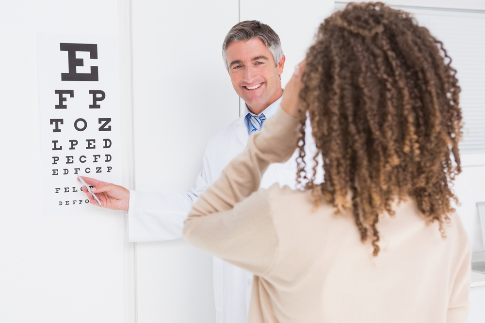
This is the part of the eye exam that most patients recognize. You’ll sit in front of a chart and read letters of decreasing size, first with one eye covered and then the other. The results are expressed as a fraction, with 20/20 being the standard benchmark for clear vision at 20 feet. Visual acuity testing tells your doctor how well your eyes are performing at that moment, but it’s a starting point, not a conclusion. A patient can pass a visual acuity test and still have significant eye disease developing beneath the surface.
2. Refractive Error
After visual acuity, your doctor will assess whether your eyes have a refractive error, meaning light isn’t focusing correctly on the retina. Nearsightedness, farsightedness, and astigmatism are all refractive errors.
Using a combination of instruments, including a phoropter (the device where you’re asked “which is clearer, one or two?”), your doctor determines the precise lens correction your eyes need. This is where your glasses or contact lens prescription comes from.
For patients considering vision correction surgery, a detailed refractive evaluation at Williamson Eye Center is also the foundation of any LASIK or PRK candidacy assessment.
3. Eye Pressure (Tonometry)
Elevated intraocular pressure is the leading risk factor for glaucoma, a condition that destroys the optic nerve without any pain or warning symptoms.
Tonometry measures the pressure inside your eyes, typically using a small puff of air or a gentle probe against the surface of the eye. Normal pressure falls within a specific range, and readings outside that range prompt further testing.
Because glaucoma causes irreversible vision loss before most patients notice anything is wrong, catching elevated pressure early is one of the most important things a routine exam can do.
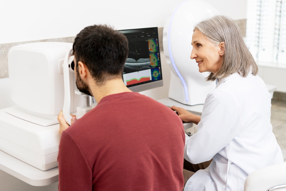
4. Optic Nerve and Retina (Dilated Exam)
Dilation is the part of the exam that makes your eyes sensitive to light for a few hours afterward, and it’s also the part that gives your doctor the clearest view of what’s happening inside your eye. Eye drops are used to widen the pupil so the doctor can examine the optic nerve head, the macula, the peripheral retina, and the blood vessels that supply them. This is where signs of macular degeneration, retinal tears, and glaucomatous nerve damage become visible, along with early diabetic retinopathy, which produces no symptoms in its earliest stages. That absence of symptoms is precisely why the dilated exam is so valuable.
5. Eye Muscle Function and Alignment
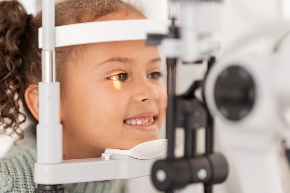
Your two eyes need to work together precisely. When they don’t, the result can be double vision, eye strain, headaches, or difficulty reading. Your doctor will test how your eyes move through their full range of motion, how well they track a moving target, and whether they point in the same direction at both near and far distances.
In children, this part of the exam is especially important. Conditions like amblyopia (lazy eye) and strabismus (crossed eyes) are far easier to treat when identified early, and they won’t always be obvious to a parent or even to the child.
6. Color Vision and Peripheral Vision
Color vision testing checks for deficiencies in how your eyes distinguish between certain colors, most commonly red and green. While color blindness is usually inherited and present from birth, changes in color perception later in life can signal retinal or optic nerve problems. Visual field testing evaluates your side vision, which is often the first type of vision affected by glaucoma and certain neurological conditions. These tests are quick and add meaningful diagnostic information to the overall picture your doctor is building.
7. Lens and Cornea Evaluation
Using a slit lamp, a specialized microscope with a bright narrow beam of light, your doctor examines the structures at the front of the eye in detail.
The cornea is evaluated for clarity, irregular curvature, and early signs of keratoconus, a progressive thinning of the cornea that distorts vision over time. The lens is examined for early cataract formation as well. Cataracts develop gradually, and patients often adapt to the slow change in their vision without realizing how much clarity they’ve lost. Identifying lens changes early gives your doctor the ability to monitor progression and plan treatment before vision is significantly compromised.
8. Dry Eye Assessment
Dry eye disease is one of the most underdiagnosed conditions in eye care. During your exam, your doctor will assess the quality and quantity of your tear film, the stability of the tear layer across the surface of your eye, and the health of the meibomian glands responsible for producing the oil that keeps tears from evaporating.
Many patients attribute their symptoms, including fluctuating vision, burning, redness, or the sensation of something in the eye, to allergies or screen fatigue. Identifying dry eye disease as the actual cause opens the door to effective treatment rather than temporary relief.
9. Your Overall Health Picture
The eyes are the only place in the body where blood vessels can be viewed directly without imaging equipment. That makes them a surprisingly informative window into your general health.
During a dilated exam, your doctor can spot signs of diabetes, high blood pressure, high cholesterol, and autoimmune disease, sometimes before a patient has received any diagnosis from their primary care physician. For patients already managing diabetes, annual diabetic eye care exams are particularly important, as retinal damage from blood sugar fluctuations can begin well before vision changes are noticeable.
A comprehensive eye exam typically takes 45 minutes to an hour, and the information it provides goes far beyond a glasses prescription. The conditions that cause the most permanent vision loss, including glaucoma, diabetic eye disease, and macular degeneration, are all most treatable when caught early, which means the exam itself is one of the most effective tools available for protecting your long-term vision.
Ready to see what a thorough eye exam can reveal? Schedule an appointment at Williamson Eye Center in Baton Rouge, LA.

